T cell stimulation for flow cytometry analysis
Activation and proliferation protocols provide an effective method to determine immunocompetence and cell reactivity. However choosing the right stimulation protocol and knowing when to use it may vary depending on what cell type you are starting with and the result you need.

To help you we have grouped a few simple protocols for T cells, with some background information so you can choose the right method. These protocols are useful when you need to measure activation markers (Figure 1), cytokine release, receptor expression and proliferation responses (Figure 2). It can also be used as a positive control for your experiment. The length of time in culture prior to analysis will determine what can be effectively measured. Activation markers are visible after a few hours whereas proliferation can take up to five days. Although these protocols will allow you to assess reactivity they will not give you information about specific effector function nor allow you to identify specific T cell subsets.
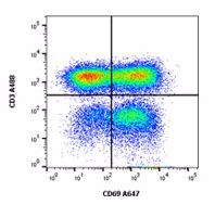
Fig. 1. PBMCs extracted from human blood were stimulated with PHA and LPS for 48 hrs. Activated T cells are seen by upregulated CD69 (MCA2806A647) expression on the CD3 (MCA463A488) positive population. Cells were gated on lymphocytes in the presence of Human Seroblock (BUF070A). Data acquired on the ZE5™ Cell Analyzer.
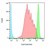
Fig. 2. Human PBLs were stained with CytoTrack Red 628/643 Cell Proliferation Assay Kit (1351205) and stimulated for five days with PHA. Cells that have proliferated show a reduction in the amount of dye with each cell division. Six peaks corresponding to each division are visible in this experiment. You can see stimulated cells (in red), labeled cells but unstimulated (in green) and unlabelled cells (in blue). Data acquired on the ZE5 Cell Analyzer.