In addition to the broad range of flow cytometry validated antibodies we offer for immunology, cancer and veterinary science, we also have supporting reagents that will optimize staining and sample preparation.

To help you use these reagents, we have created a free poster, available to download or order, which takes you through the important stages you should follow to design a successful flow cytometry experiment.
Buffers
We have a range of buffers suitable for use in cell surface and intracellular antibody staining for flow cytometry and other applications.
Leucoperm™– cell permeabilization reagent for intracellular staining
Leucoperm reagent allows intracellular antigen analysis with the same ease as surface antigens. Structures such as cytoplasmic or nuclear enzymes, oncoproteins, cytokines and cytoplasmic localizations of established membrane molecules can be stained with antibodies. Leucoperm allows you to fix and permeabilize cells in suspension whilst retaining their morphology.
Staining buffer – bovine calf serum containing staining buffer
Our staining buffer has been optimized for use when staining both live and fixed cells with antibodies. Suitable for use with both conjugated and unconjugated antibodies and can be used as an antibody dilution media and a wash buffer. The addition of bovine calf serum reduces background staining to target cells and contains sodium azide to minimize receptor capping.
Fixation buffer – paraformaldehyde containing fixation buffer
Essential for intracellular staining prior to permeabilization, fixation buffer is a paraformaldehyde based fixative suitable for use on most cell types.
ICS perm wash buffer (10x) – permeabilization and wash buffer for intracellular staining
Access to intracellular targets such as cytokines and cytoplasmic proteins requires permeabilization of the cell membrane after fixation. Our permeabilization buffer has minimal effect on cell morphology and is supplied as a 10x solution for dilution in water. It can also be used as a wash buffer during intracellular staining to help minimize background staining.
Activation Reagents
We offer two cell activation reagents, with and without a protein transport inhibitor, containing optimized concentrations of phorbol 12-myristate-13-acetate (PMA) and ionomycin, which can be used to activate your cells of interest.
Cell Stimulation Reagent (without Brefeldin A)
An optimized mixture of PMA and ionomycin in DMSO without a protein transport inhibitor, suitable for activating cells and for stimulating cytokine secretion. Available as a 500x solution in DMSO. For intracellular cytokine staining we recommend adding either Brefeldin A or Monensin or using our Cell Stimulation Reagent with Brefeldin A to allow accumulation of cytokines in the cell.
Cell Stimulation Reagent (with Brefeldin A)
An optimized mixture of PMA and ionomycin in DMSO with the protein transport inhibitor Brefeldin A, suitable for activating cells and increasing cytokine production. Brefeldin A allows cytokines to accumulate within the cell allowing detection by intracellular fluorescent staining.
Protein transport inhibitors
We offer two protein transport inhibitors which can be used to enhance intracellular cytokine staining. As cytokines and chemokines are usually secreted, use of these inhibitors, trapping the proteins within the cell, leads to a build-up of cytokines in the golgi apparatus and endoplasmic reticulum. The choice and culture conditions for these inhibitors may need optimizing depending on the species and cytokines being stained.
This inhibitor acts by interfering with the Na+/H+ transport in golgi apparatus transmembranes. Available as a 1000x solution in 70% ethanol.
This inhibitor acts by redistributing proteins from the golgi apparatus to the endoplasmic reticulum. Available as a 1000x solution in DMSO.
Fc blocks
Fc receptors are found on monocytes, macrophages, dendritic cells and B cells. As the name suggests they bind antibodies via their constant Fc domain rather than the antigen specific Fab domain. This can lead to false positives. In order to prevent this type of binding, blocking agents have been developed to ensure that only antigen specific binding is observed.
Mouse Seroblock – for blocking Fc receptors on mouse cells
Human Seroblock – for blocking Fc receptors on human cells
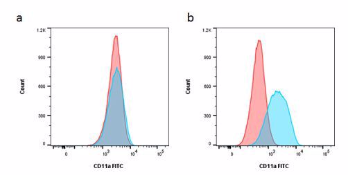
Fig.1. Staining of THP-1 cells with FITC labeled mouse anti-human CD11a in blue (MCA1848) or mouse IgG2a negative control:FITC red (MCA929F) in the absence (a) and presence (b) of human SeroBlock (BUF070A).
Red cell lysing buffer – Erythrolyse is available as a 10x solution stable for 1 year at room temperature. It is suitable for the lysing of red blood cells in human, rat and mouse blood.
Viability dyes – we have easy-to-use cell viability assays in a range of excitation and emission wavelengths, suitable for most multi-color flow cytometry. These include DNA binding dyes such as PI, 7-AAD and DAPI and our protein binding VivaFixTM dyes which are fixable to allow analysis at a later date.
Proliferation dyes – our cell proliferation assays include dyes, TUNEL assay kits and BrdU antibodies to give you flexibility in your experiments. They are also available in a range of excitation and emission wavelengths for your convenience.
Isotype controls – determine the level of background surface staining due to non-specific antibody or fluorophore binding. We have isotype controls for mouse, rat, hamster and goat antibodies.
When performing multicolor flow cytometry, there are several important steps you must consider. Careful sample preparation, Fc blocking, inclusion of a live/dead dye, doublet exclusion and using the appropriate controls are all important. For more information watch our flow cytometry webinar Optimize your Flow Cytometry or visit our application resources page.