The ZOE™ Fluorescent Cell Imager eliminates the complexities of cell imaging associated with traditional microscopes. This fluorescence imaging system combines the ease of use of a personal tablet with the power of an inverted microscope.
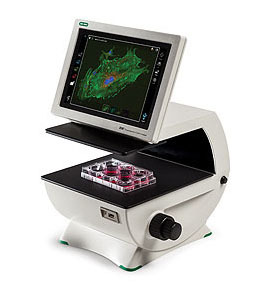
An Android-based platform, the ZOE™ Cell Imager uses an intuitive touch-screen interface to control the brightfield, three fluorescence channels, and the integrated digital camera.
The ZOE™ Fluorescent Cell Imager is a complete digital imaging system, allowing users to view samples, capture and store images, and create multicolor overlays. Thanks to the built-in light shield, the ZOE™ Cell Imager does not require a darkroom for fluorescence imaging.
- Simplified cell imaging — the intuitive touch-screen interface allows users to view cells, capture images, and create multichannel merges with minimal training
- Flexible operation — brightfield and three fluorescence channels enable use for routine cell culture applications and more sophisticated imaging applications
- Fluorescence imaging at your bench — light shield permits fluorescence imaging in ambient light
- Robust construction — fully integrated system with long-life LEDs, ready for intensive daily use
- LED light sources — thousands of hours of illumination that are instantly ready after power-on
- Large viewing area — the motorized stage and wide field of view allow you to see more of your sample, faster
- Small footprint — compact size accommodates crowded lab benches
Applications of the ZOE Fluorescent Cell Imager
Use the ZOE Cell Imager to check/screen samples prior to high-content analysis (HCA), high-throughput screening (HCS), confocal imaging, or fluorescence-activated cell sorting (FACS). With a brightfield and three fluorescent channels, the ZOE Cell Imager has all the features needed for daily cell culture work as well as fluorescent applications:
- Visual estimation of cell confluency
- Observation of general cell health and morphology
- Cell growth and proliferation monitoring
- Capturing cell images (with or without fluorescent labels)
- Visualization of expressed fluorescent proteins
- Immunofluorescent protein localization
- Estimation of transfection efficiency
Illumination Light Sources
- Blue channel uses a UV LED
- Green channel uses a blue LED
- Red channel uses a green LED
- Brightfield channel uses a ring of multiple green LEDs for reduced chromatic aberration