Use ReadiLink Antibody Conjugation Kits to quickly conjugate microscale volumes of antibody (50-100 µg) in two easy steps with the unique chemistry of ReadiLink Dyes. Fluorophores are available that are excited by violet all the way to infrared with a range of emission spectra to easily fit into your multicolor flow cytometry experiments, giving you greater choice.
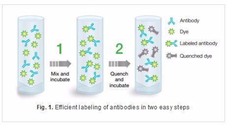
The unique chemistry of Readilink Dyes allows you to quickly label your antibody in just two easy steps, shown in Figure 1.
Each Readilink Dye Kit includes succinimidyl esters that selectively react with primary amines to form a stable carboxamide bond. This bond ensures no dissociation between the fluorophore and antibody which may lead to unbound fluorophore in your experiments.
Readilink Antibody Conjugation Kits key benefits
-
Saving you time: As no purification column required
-
Minimal hands on time: Easy protocol doesn’t involve complicated chemistry, allowing you to perform other experiments
-
Ready-to-use: No fluorophore quenching required
-
Versatile: Available in 10 unique fluorophores with a wide range of excitation wavelengths – giving you greater flexibility in your multicolor flow cytometry and cell sorting experiments as well as other applications such as western blot and ELISA
Readilink Conjugation Kits allow you to label your primary antibody quickly avoiding the use of secondary antibodies and can help improve your flexibility in building panels. Purified CD45RA was labeled with Readlink 405/454 1351011 and human peripheral blood stained in combination with CD3 FITC (MCA463F) and CD4 (MCA1267PE). When compared to a Pacific Blue™ directly conjugated CD45RA (MCA88PB) antibody the staining was comparable allowing identification of naïve CD4 positive T cells.
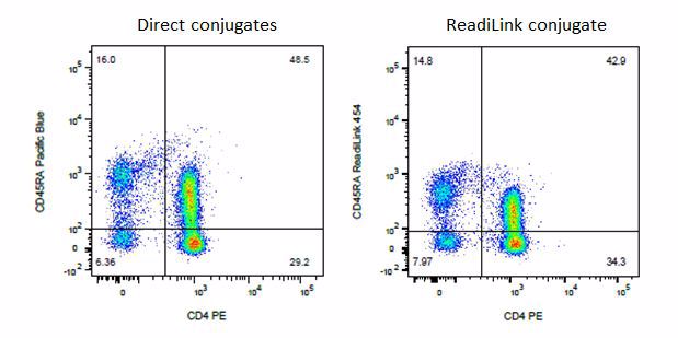
Fig.2. Purified CD45RA (MCA88) was labeled with Readilink 405/454 and used to stain human peripheral blood in combination with CD4 PE (MCA1267PE). Alternatively directly labeled CD45RA Pacific Blue (MCA88PB) was used. The staining shown is lymphocytes gated on the CD3 positive population. CD4 and CD45RA positive naïve T cells can be identified in both plots.